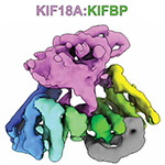
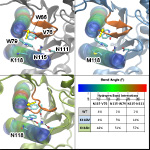
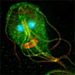
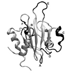
Kinesin-binding protein remodels the kinesin motor to prevent microtubule binding
A. L. Solon, Z. Tan, K. L. Schutt, L. Jepsen, S. E. Haynes, A. I. Nesvizhskii, D. Sept, J. Stumpff, R. Ohi and M. A. Cianfrocco
Sci Adv 7 (47) eabj9812, 2021.
Parthenolide Destabilizes Microtubules by Covalently Modifying Tubulin
Curr Biol 31 (4) 900-907 e6, 2021.
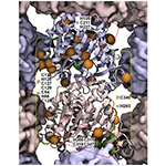
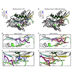
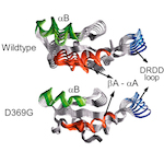
Pathogenic mutations in the kinesin-3 motor KIF1A diminish force generation and movement through allosteric mechanisms
J Cell Biol 220 (4) 2021.
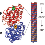
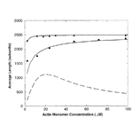
Effects of Nucleotide and End-Dependent Actin Conformations on Polymerization
Biophys J 119 (9) 1800-1810, 2020.
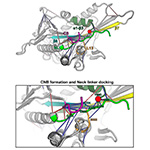
Neck linker docking is critical for Kinesin-1 force generation in cells but at a cost to motor speed and processivity
Elife 8 2019.
The Structure and Dynamics of C. elegans Tubulin Reveals the Mechanistic Basis of Microtubule Growth
Dev Cell 2018.
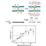
Frequency-Based Analysis of Gramicidin A Nanopores Enabling Detection of Small Molecules with Picomolar Sensitivity
Anal Chem 90 (3) 1635-1642, 2018.
Cytokine receptor-Eb1 interaction couples cell polarity and fate during asymmetric cell division
Elife 7 2018.

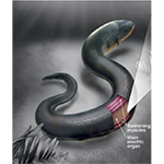
An electric-eel-inspired soft power source from stacked hydrogels
Nature 552 (7684) 214-+, 2017.
Real-time shape approximation and fingerprinting of single proteins using a nanopore
Nat Nanotechnol 12 (4) 360-367, 2017.
14-3-3 Regulates Actin Filament Formation in the Deep-Branching Eukaryote Giardia lamblia
mSphere 2 (5) 2017.
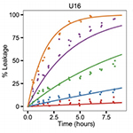
Hybrid Lipids Inspired by Extremophiles and Eukaryotes Afford Serum-Stable Membranes with Low Leakage
Chemistry 23 (28) 6757-6762, 2017.
Mechanisms of kinetic stabilization by the drugs paclitaxel and vinblastine
Mol Biol Cell 28 (9) 1238-1257, 2017.

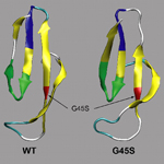
Two Deafness-Causing Actin Mutations (DFNA20/26) Have Allosteric Effects on the Actin Structure
Biophys J 111 (2) 323-32, 2016.
Effects of Lipid Tethering in Extremophile-Inspired Membranes on H+/OH- Flux at Room Temperature
Biophysical Journal 110 (11) 2430-2440, 2016.
Novel alpha-tubulin mutation disrupts neural development and tubulin proteostasis
Developmental Biology 409 (2) 406-419, 2016.
Hydrogel-assisted functional reconstitution of human P-glycoprotein (ABCB1) in giant liposomes
Biochim Biophys Acta 1848 (2) 643-53, 2015.

Modeling large-scale dynamic processes in the cell: polarization, waves, and division
Quarterly Reviews of Biophysics 47 (3) 221-248, 2014.
A method for multiprotein assembly in cells reveals independent action of kinesins in complex
Journal of Cell Biology 207 (3) 393-406, 2014.
Assessing the barriers to image-guided drug delivery
Wiley Interdisciplinary Reviews-Nanomedicine and Nanobiotechnology 6 (1) 1-14, 2014.

Capping protein regulators fine-tune actin assembly dynamics
Nat Rev Mol Cell Biol 15 (10) 677-89, 2014.
Genome-wide Analysis Reveals Novel and Discrete Functions for Tubulin Carboxy-Terminal Tails
Current Biology 24 (12) 1295-1303, 2014.
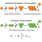
The unusual dynamics of parasite actin result from isodesmic polymerization
Nat Commun 4 2285, 2013.
Properties of Microtubules with Isotropic and Anisotropic Mechanics
Cellular and Molecular Bioengineering 6 (4) 361-368, 2013.
Multisite ion models that improve coordination and free energy calculations in molecular dynamics simulations
J. Chem. Theor. Comp. 9 (8) 3538-42, 2013.
A model for the interfacial kinetics of phospholipase D activity on long-chain lipids
Biophys J 105 (1) 146-53, 2013.

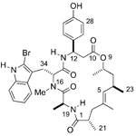
Synthetic chondramide A analogues stabilize filamentous actin and block invasion by Toxoplasma gondii
J Nat Prod 76 (9) 1565-72, 2013.
Mechanical properties of doubly stabilized microtubule filaments
Biophys J 104 (7) 1517-28, 2013.
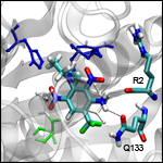
Carboxyl Group Footprinting Mass Spectrometry and Molecular Dynamics Identify Key Interactions in the HER2-HER3 Receptor Tyrosine Kinase Interface
J Biol Chem 288 (35) 25254-64, 2013.

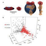
Multivariate analyses of amyloid-beta oligomer populations indicate a connection between pore formation and cytotoxicity
PLoS One 7 (10) e47261, 2012.

Single-particle characterization of abeta oligomers in solution
ACS Nano 6 (7) 5909-19, 2012.

Mechanism for CARMIL Protein Inhibition of Heterodimeric Actin-capping Protein
Journal of Biological Chemistry 287 (19) 15251-15262, 2012.
Perturbations in Microtubule Mechanics from Tubulin Preparation
Cellular and Molecular Bioengineering 5 (2) 227-238, 2012.
Synthesis of Chondramide A Analogues with Modified beta-Tyrosine and Their Biological Evaluation
Chem. Eur. J. 17 (47) 13349-57, 2011.
Drug-like Leads for Steric Discrimination between Substrate and Inhibitors of Human Acetylcholinesterase
Chem Biol Drug Design 78 (4) 495-504, 2011.

Evolutionarily divergent, unstable filamentous actin is essential for gliding motility in apicomplexan parasites
PLoS Pathog 7 (10) e1002280, 2011.
Pharmacokinetic modeling of tumor bioluminescence implicates efflux, and not influx, as the bigger hurdle in cancer drug therapy
Cancer Research 71 (3) 686-92, 2011.
An actin cytoskeleton with evolutionarily conserved functions in the absence of canonical actin-binding proteins
Proc. Natl. Acad. Sci. 108 (15) 6151-6, 2011.
An Epilepsy/Dyskinesia-Associated Mutation Enhances BK Channel Activation by Potentiating Ca2+ Sensing
Neuron 66 (6) 871-883, 2010.
Microtubule elasticity: connecting all-atom simulations with continuum mechanics
Phys Rev Lett 104 (1) 018101, 2010.

F-actin structure destabilization and DNase I binding loop: fluctuations mutational cross-linking and electron microscopy analysis of loop states and effects on F-actin
J Mol Biol 395 (3) 544-57, 2010.

Dinitroaniline activity in Toxoplasma gondii expressing wild-type or mutant alpha-tubulin
Antimicrob Agents Chemother 54 (4) 1453-60, 2010.
Overlapping and distinct functions for cofilin, coronin, and Aip1 in actin dynamics in vivo
J Cell Sci 123 (Pt 8) 1329-42, 2010.
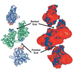
The interaction of capping protein with the barbed end of the actin filament
J Mol Biol 404 (5) 794-802, 2010.
An actin-filament-binding interface on the Arp2/3 complex is critical for nucleation and branch stability
Proc Natl Acad Sci U S A 2010.
The Kinetics of Cooperative Cofilin Binding Reveals Two States of the Cofilin-Actin Filament
Biophysical Journal 98 (9) 1893-1901, 2010.
A multiscale model linking ion-channel molecular dynamics and electrostatics to the cardiac action potential
Proc Natl Acad Sci U S A 106 (27) 11102-6, 2009.
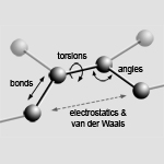
The Basic Concepts of Molecular Modeling
Methods in Enzymology: Computer Methods, Part B 467 307-334, 2009.
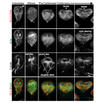
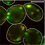
Neurodegeneration mutations in dynactin impair dynein-dependent nuclear migration
Proc Natl Acad Sci U S A 106 (13) 5147-52, 2009.
A22 disrupts the bacterial actin cytoskeleton by directly binding and inducing a low-affinity state in MreB
Biochemistry 48 (22) 4852-7, 2009.

Nanoparticle pharmacokinetic profiling in vivo using magnetic resonance imaging
Magn Reson Med 60 (6) 1353-61, 2008.

Taxol allosterically alters the dynamics of the tubulin dimer and increases the flexibility of microtubules
Biophys J 95 (7) 3252-8, 2008.
Mapping the cofilin binding site on yeast G-actin by chemical cross-linking
J Mol Biol 377 (2) 395-409, 2008.

Effects of solution crowding on actin polymerization reveal the energetic basis for nucleotide-dependent filament stability
J Mol Biol 378 (3) 540-50, 2008.
New insights into the mechanism and regulation of actin capping protein
Int Rev Cell Mol Biol 267 183-206, 2008.
Nucleotide effects on the structure and dynamics of actin
Biophys J 93 (4) 1277-83, 2007.
First-contact time to a patch in a multidimensional potential well
Phys Rev E Stat Nonlin Soft Matter Phys 76 (2 Pt 1) 021911, 2007.
Mutations in alpha-tubulin confer dinitroaniline resistance at a cost to microtubule function
Mol Biol Cell 18 (12) 4711-20, 2007.
Structure/function analysis of the interaction of phosphatidylinositol 4,5-bisphosphate with actin-capping protein: implications for how capping protein binds the actin filament
J Biol Chem 282 (8) 5871-9, 2007.
Energetics and dynamics of constrained actin filament bundling
Biophys J 90 (12) 4295-304, 2006.
Unusual kinetic and structural properties control rapid assembly and turnover of actin in the parasite Toxoplasma gondii
Mol Biol Cell 17 (2) 895-906, 2006.
Binding and interaction of dinitroanilines with apicomplexan and kinetoplastid alpha-tubulin
J Med Chem 49 (17) 5226-31, 2006.
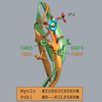
Myo1c binds phosphoinositides through a putative pleckstrin homology domain
Mol Biol Cell 17 (11) 4856-65, 2006.
A genetic dissection of Aip1p’s interactions leads to a model for Aip1p-cofilin cooperative activities
Mol Biol Cell 17 (4) 1971-84, 2006.
Binding of myotrophin/V-1 to actin-capping protein: implications for how capping protein binds to the filament barbed end
J Biol Chem 281 (41) 31021-30, 2006.
The NH2 terminus of RCK1 domain regulates Ca2+-dependent BK(Ca) channel gating
J Gen Physiol 126 (3) 227-41, 2005.
Localization of the antimitotic peptide and depsipeptide binding site on beta-tubulin
Biochemistry 43 (44) 13955-62, 2004.

The physical basis of microtubule structure and stability
Protein Sci 12 (10) 2257-61, 2003.
Thermodynamics and kinetics of actin filament nucleation
Biophys J 81 (2) 667-74, 2001.
Electrostatics of nanosystems: application to microtubules and the ribosome
Proc Natl Acad Sci U S A 98 (18) 10037-41, 2001.
Kinetic mechanism of end-to-end annealing of actin filaments
J Mol Biol 312 (4) 721-30, 2001.

Commitment to folded and aggregated states occurs late in interleukin-1 beta folding
Biochemistry 39 (50) 15633-42, 2000.
Annealing accounts for the length of actin filaments formed by spontaneous polymerization
Biophys J 77 (6) 2911-9, 1999.
A chemical kinetics model for microtubule oscillations
J Theor Biol 197 (1) 77-88, 1999.

Computer simulations of actin polymerization can explain the barbed-pointed end asymmetry
J Mol Biol 294 (5) 1181-9, 1999.
Model for spatial microtubule oscillations
Phys Rev E Stat Phys Plasmas Fluids Relat Interdiscip Topics 60 (1) 838-41, 1999.
Selected physical issues in the structure and function of microtubules
J Struct Biol 118 (2) 94-106, 1997.